DISIOR
  • Home
  • Products
    • CMF Orbital
    • Bonelogic 2.0
  • Research
  • About us
    • In Media
    • Team
    • Careers
    • Contact
  • Blog
Click to set custom HTML

Diagnosis and treatment through true 3D vision


We develop 3D medical imaging software for clinicians


​Our mission is to provide medical professionals with the diagnostic information they need to deliver perfectly-tailored treatments to every patient.
Using our software, you can segment, quantify and model 3D medical imaging data in minutes. The results from our software provide highly accurate patient-specific 3D models and clinically relevant 3D measurements that provide objective data for diagnosis, treatment planning and outcome assessment.      
Speak to an expert
 
Professor Jari Salo, MD & Specialist in Orthopaedics and Trauma
“Until today, 3D medical images have been analyzed using 2D slices, as if you were looking at the image with one eye only. The Disior method analyzes the original 3D data in a way that compares to having a full vision.”​
​
– Professor Jari Salo, MD & Specialist in Orthopedics and Trauma

Key benefits


Save Time

Experience a 95%  reduction in medical image processing times.

​Go from DICOM to 3D anatomical models and analytics in less than 5 minutes. ​ 

Be Objective

Disior's 3D anatomical analysis is developed around clinical landmarks and reference points that can be defined objectively with a high degree of repeatability.

Get Better Outcomes 

Use 3D measurements to accurately diagnose and quickly plan your patients' treatment.

Conduct large scale orthopaedic studies to improve understanding of pathological conditions and normal variation. 
"Disior's innovation leverages the full amount of 3D imagery directly to the doctor. This enables accurate and rapid diagnosis and in the long run also increases the systematic nature of treatment."
- Professor Risto Kontio, Chief Physician of Oral and Maxillofacial Surgery, Helsinki University Hospital
Picture

Introducing Bonelogic® 2.0

Become a leader in three-dimensional orthopaedics
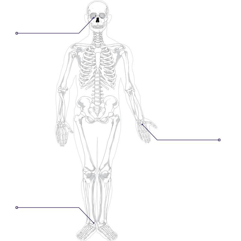
3D anatomical model of a hand and wrist and foot and ankle
We have combined our innovative Ortho Foot & Ankle and Ortho Hand & Wrist products into one easy to use 3D medical imaging software that gives you:

1) 
Fully segmented medical imaging data from CT, CBCT and WBCT in minutes.
2) 
Mathematically accurate patient-specific 3D models.
3) Objective and reliable angles and distances that describe the relationship between bones in three-dimensions. ​
​
All this makes Bonelogic 2.0 excellent for assessing common injuries and diseases like flat foot deformity, osteoarthritis, ankle instability and radius fractures

Book a call with an ExperT
An in depth call with one of our experts will typically last 45 mins.
In that time we can discuss your current analysis workflow, demonstrate a product of your choosing and discuss pricing.

Trusted By

Picture
Picture
Picture
Picture
Picture
Picture
Picture
Picture
Picture
Picture
Picture
Picture

Our 3D Medical Imaging Software Products

Medical imaging software for 3D foot & ankle analysis

Foot & Ankle analysis -
Bonelogic
® 2.0

Automatic analysis of the foot and ankle anatomy as well as radiographic parameters.
WATCH TUTORIAL
Medical imaging software for orbital volume and fracture analysis

Orbital fracture analysis -
Bonelogic
® CMF Orbital

Automatic analysis of the shape and size of orbital fractures, and virtually reconstruct.
​

WATCH TUTORIAL
Medical imaging software for 3D hand & wrist analysis

Hand & Wrist analysis -
Bonelogic
® 2.0

Automatic analysis of the hand and wrist anatomy as well as radiographic parameters.
WATCH TUTORIAL

Research Highlights

Ceramics International
​

Lowe et al.
​FEA evaluation of material stiffness changes for a polymer assisted 3D polycaprolactone/β-tricalcium phosphate scaffold in a mandibular defect reconstruction model

read more

Journal of Hand Surgery

​Suojärvi et al.
Radiographic measurements of the normal distal radius: reliability of computer-aided CT versus physicians' radiograph interpretation

read more

Foot & Ankle International

Kvarda et al.
3D Assessment in post-traumatic ankle osteoarthritis
​
​
READ MORE

In Partnership With

Picture
Picture
Picture
Picture

Our Story
We believe that doctors should have the best possible tools to help them treat their patients. That's why we bring advanced medical imaging software to doctors' day-to-day work. As engineers use mathematic modelling in industry to help with mechanics, motion and strength calculations - so can the physicians in clinical work.

The story of Disior starts back in 2016 when we decided to re-purpose a software used in mobile phone development, after seeing the potential benefits it could have in medicine. As a result of years of working together with medical doctors, researchers and professors, Disior has developed tools which maximize the benefit of today's 3D medical images for better diagnostics and treatment planning.

With Disior - Doctors get all the data they need from medical images - only faster, easier and with more accuracy.


Today, our software is being used in Maxillofacial and Orthopedic surgery all over the world.


MEET THE TEAM

See the future with your own eyes

Contact one of our in-house experts to discuss your medical image processing needs, see Bonelogic® in action or request a 30-day free trial.
Talk to us
Analyzed 3D model of the foot and ankle
Automatic orbital fracture detection
Analyzed 3D model of the hand and wrist
Picture

Diagnosis and treatment through true 3D vision


Are you interested in our products? Contact our sales team: sales@disior.com 
​

​
Do you need help?
Contact our support team: 
support@disior.com


Follow our journey at:
​
Copyright © 2021 Disior Ltd. All Rights Reserved Privacy Notice

Picture
  • Home
  • Products
    • CMF Orbital
    • Bonelogic 2.0
  • Research
  • About us
    • In Media
    • Team
    • Careers
    • Contact
  • Blog