“It was one of these subtle cases and I needed to get an accurate 3D model and distance map to see how to approach this patients’ treatment." - François Lintz, MD. 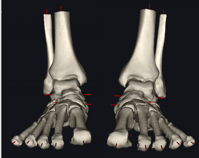 The accurate 3D models, revealed a very subtle but asymmetrical TMT1 arthritis on the left foot, where the metatarsal had slipped on the cuneiform (Figure 1). 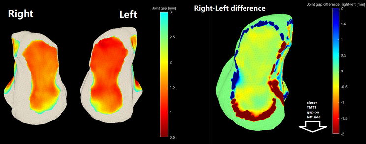 Figure 2. Right-left comparison of TMT1 joint space. By comparing the joint spacing between the left and right TMT1 you can see the subtle narrowing of the left TMT1 joint indicative of arthritis (Figure 2). Figure 3. Video of the distance mapping of the Lisfranc area. Distance mapping of the Lisfranc region (Figure 3) showed subluxation and approximation in the dorsal aspect of the joint explaining the patient's pain. These insights allowed Dr Lintz to choose the most appropriate treatment, firstly a pair of supporting insoles, then a TMT1 fusion. “This case clearly illustrates why Disior’s software is a game changer for common foot problems." |
||||||||


| |
| | ||
Disior™ Bonelogic™ was built specifically to help clinicians make diagnoses and plan patient treatment. Bonelogic™ takes DICOMs from CT, CBCT or WBCT and generates accurate models and analytics that describe the relationships between bones, like those used in this case study.
If you're interested in learning how Disior's products can aid in your clinical practice, talk to one of our experts. In a 30 minute meeting we can talk through your current image analysis workflow and discuss what we can do to improve patient outcomes together.
If you're interested in learning how Disior's products can aid in your clinical practice, talk to one of our experts. In a 30 minute meeting we can talk through your current image analysis workflow and discuss what we can do to improve patient outcomes together.
Comments are closed.
Stories from the clinic
These are written in collaboration with our customers and clinical advisors.
Archives
November 2021
October 2021
July 2021
May 2021
 RSS Feed