
|
The fundamental promise of advanced medical imaging techniques like CBCT and WBCT is improving patient care. With better patient care being achieved through automation of image analysis, increased clinical knowledge and the development of diagnostic metrics. Within extremity orthopedics 3D X-ray techniques are aiding diagnosis and treatment of common foot and ankle conditions like bunions and flat foot. The development and adoption of tools that can automate analysis of 3D medical images is a priority for this community. As automating analysis means removing barriers to both large-scale research projects (that can define normal ranges of bone alignment and objective metrics for defining pathologies and injuries) and use of 3D analytics within clinical practice. Increased collaboration between clinical orthopedic researchers and software developers is vital to achieving the goal of improved patient care.
Dr. Alberto Leardini is author of >190 scientific papers on extremity biomechanics and image analysis, mainly of the foot and ankle. He heads the Movement Analysis Lab group at IOR and is a prominent member of the WBCT society. The first results of the collaboration between Dr Leardini’s group and Disior™, which started at AOFAS 2019, are being presented at i-FAB Congress this April. A similar presentation was also given at the January WBCT virtual meeting, which was covered on our blog and is available to watch on FootInnovate.
Disior™ Bonelogic™ was built specifically to overcome some of issues of automation discussed here. Bonelogic™ takes DICOMs from CT, CBCT or WBCT and generates accurate models and analytics that describe the relationships between bones, in minutes. For example, across the 2600-foot datasets analyzed with Bonelogic™ in 2020 the average time taken for the models and analytics to be generated was 3.1 minutes. If you're interested in learning how Disior's products can aid in your research activities or clinical practice, talk to one of our experts. In a 45 minute meeting we can talk through your current image analysis workflow and discuss what we can do to improve patient outcomes together. 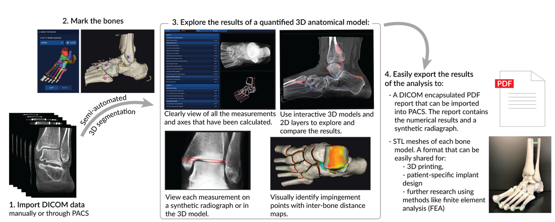 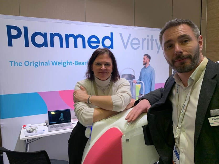 On February 5th, Disior™’s class II medical imaging software device for treatment and surgical planning in the field of orthopedics (foot & ankle and hand & wrist) Bonelogic™ (Bonelogic 2.0) received 510(k) clearance from the US FDA. Within the Planmed-Disior distribution agreement Bonelogic is paired with Planmed’s Verity. The Verity is a low-dose extremity CT scanner which provides 3D images of extremities like the hand & wrist and the foot & ankle. It was the first device in the market able to provide 3D images of the foot & ankle region under natural loading conditions, an imaging technique now commonly termed weight-bearing CT (WBCT). WBCT scanners like the Verity alongside automated medical imaging software like Bonelogic are considered are to be at the forefront of medical innovations for extremity orthopedics. Within the foot & ankle complex, WBCT is a proven tool in both European and American orthopedic clinics for the following common conditions:
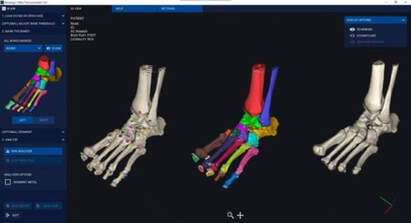
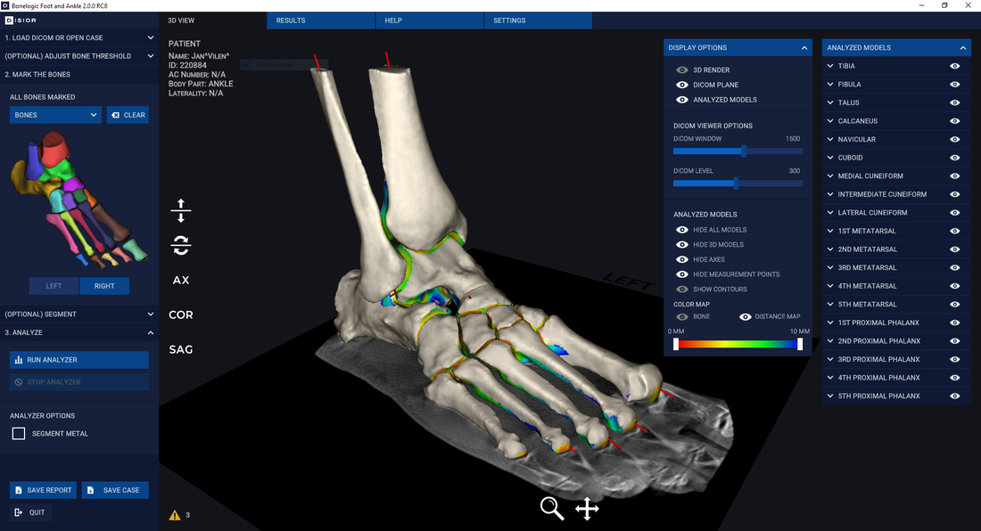
Bonelogic™ allows for complete orthopedic characterization of the foot & ankle, as well as the hand & wrist. The software provides mathematical models of a patients’ anatomy and metrics that describe the relationship between bones, which are essential to understanding the degree of pathology or trauma. “The technology within Bonelogic™ means that this type of analysis is completed in minutes. Whereas, in most other medical imaging software it would take hours. Therefore, with Bonelogic™ the barriers to clinical adoption of 3D analytics are removed.” Anna-Maria Henell, CEO, Disior. The magic of combining the distribution of the Verity extremity CT and Bonleogic is that clinical workflows for common hand & wrist and foot & ankle conditions can be optimized. Reducing the workload of radiologists and clinicians.
Disior Oy and Planmed
Disior Oy's mission is to provide medical professionals with the diagnostic information they need to deliver perfectly-tailored treatments to every patient. To do this Disior develops novel ways to automate the analysis of bones and soft tissue, linking medical doctors with engineers and mathematicians. The company owners include both leading orthopedic and maxillofacial surgeons as well as technology visionaries. Planmed Oy dedicates its effort to the development, manufacturing and marketing of advanced imaging equipment and accessories that provide a unique combination of image quality and ease of use for medical imaging professionals. The company offers products for mammography and orthopedic imaging that are well-known for imaging performance, user-friendliness and good ergonomics. Since 1989, Planmed systems have provided tools for healthcare professionals in over 70 countries worldwide. Planmed Oy is part of the Finnish Planmeca Group, which operates in the field of health care technology. |


 RSS Feed

Transforming Treatments
|
Follow our journey at:
Copyright © 2023 Disior™ Ltd. All Rights Reserved Privacy Notice License Agreement Disior™ a Paragon 28® Company |

|