DISIOR
  • Home
  • Available Modules
    • Foot & Ankle
    • Planning
    • Hand & Wrist
    • CMF Orbital
  • Clinical Research
  • Expert Cases
  • News
    • Press
    • Blog
  • About us
    • Contact Us
    • Paragon 28®
    • Careers

Collaboration is key: Disior™ partnership with Movement Analysis Lab at the Istituto Ortopedico Rizzoli (IOR) in Bologna sees results

11/3/2021

 
The fundamental promise of advanced medical imaging techniques like CBCT and WBCT is improving patient care. With better patient care being achieved through automation of image analysis, increased clinical knowledge and the development of diagnostic metrics.

Within extremity orthopedics 3D X-ray techniques are aiding diagnosis and treatment of common foot and ankle conditions like bunions and flat foot. The development and adoption of tools that can automate analysis of 3D medical images is a priority for this community. As automating analysis means removing barriers to both large-scale research projects (that can define normal ranges of bone alignment and objective metrics for defining pathologies and injuries) and use of 3D analytics within clinical practice.
Increased collaboration between clinical orthopedic researchers and software developers is vital to achieving the goal of improved patient care.
 “As MedTech innovators, Disior’s products are worthless without clinical collaboration and validation, and it’s not a one-way relationship. Software development at Disior is the results of a productive symbiosis between our team of mathematicians and engineers and our medical partners, both contributing specific skills and expertise towards our common goal of improving patient care.”  - Anna-Maria Henell, Disior™ CEO.
Disior CEO Anna-Maria Henell
Anna-Maria Henell, Disior CEO
Dr. Alberto Leardini is author of >190 scientific papers on extremity biomechanics and image analysis, mainly of the foot and ankle. He heads the Movement Analysis Lab group at IOR and is a prominent member of the WBCT society. The first results of the collaboration between Dr Leardini’s group and Disior™, which started at AOFAS 2019, are being presented at i-FAB Congress this April. A similar presentation was also given at the January WBCT virtual meeting, which was covered on our blog and is available to watch on FootInnovate.
3D models of the foot and ankle generated in Bonelogic medical imaging software
The process of generating 3D anatomical models of the foot & ankle with Bonelogic. Credit- Dr. Leardini.
Picture
Example of whole foot 3D model in Bonelogic. Credit-Disior.
This study focuses on the 3D geometrical analysis of acquired adult flatfoot deformity (AAF) or progressive collapsing foot deformity (PCFD). AAF is a common deformity that requires multiple, complex and combined modifications of the foot skeletal architecture. An exact assessment of bone alignment in the foot is vital to planning and treating this condition. WBCT provides precise imagery of the foot that allows for bone alignment to be quantified before and after surgical intervention. In this study, 3D bone models were obtained using Bonelogic™ Ortho Foot and Ankle and compared with other state-of-the-art tools. Principal Component Analysis defined the anatomical reference frames for each bone model. Absolute and relative angles were calculated, in anatomical plane projections and in 3D. The results illustrate the advantage of these techniques and tools to diagnose and evaluate the clinical treatment of AAF. Concluding that the extent of a patients AAF as assessed from WBCT and 3D metrics can be quantified exactly. 
“Diagnosis and treatment of foot and ankle diseases , as well as the assessment of the treatments, must rely on accurate description of bone alignments when the foot is in weight-bearing, in support to all the traditional clinical measures. Establishing common terminology, definitions, and tools for analyzing and reporting these bone alignments in 3D is now essential. This research paper is part of a drive that will ultimately lead also to better medical understanding of the foot complex, and more informed patient care.” - Dr Leardini, IOR.
Dr Alberto Leardini, Istituto Ortopedico Rizzoli
Dr. Alberto Leardini, Head of Movement Analysis Lab at Istituto Ortopedico Rizzoli

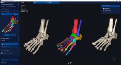
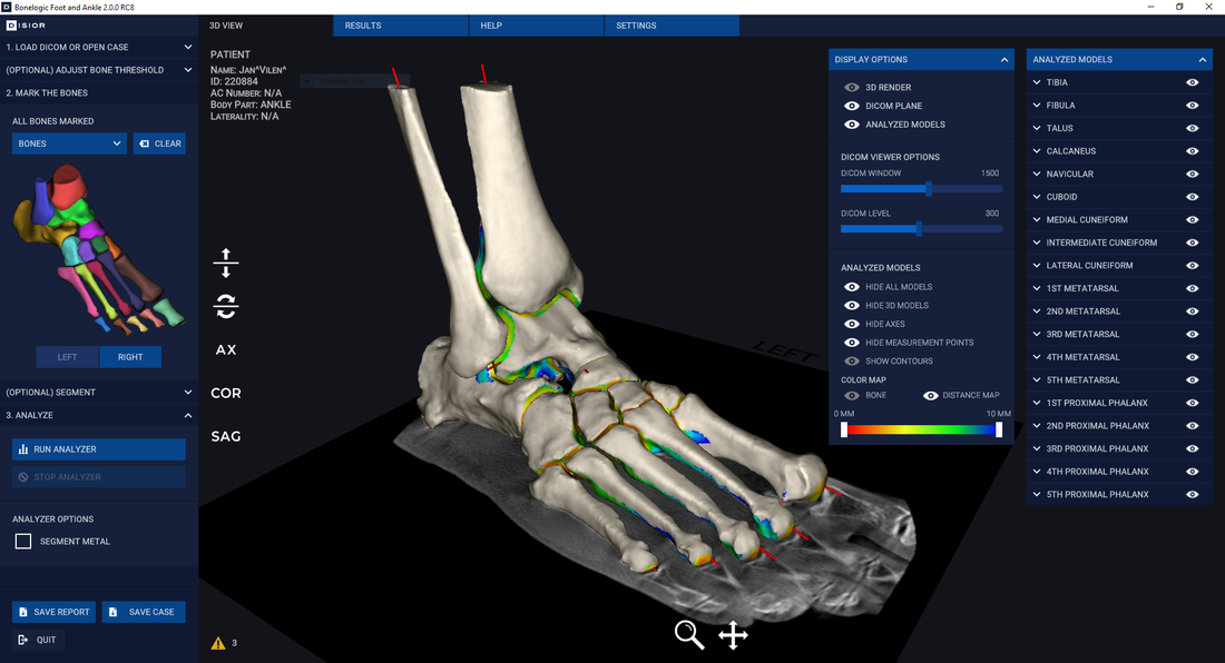
Disior™ Bonelogic™ was built specifically to overcome some of issues of automation discussed here. Bonelogic™ takes DICOMs from CT, CBCT or WBCT and generates accurate models and analytics that describe the relationships between bones, in minutes. For example, across the 2600-foot datasets analyzed with Bonelogic­™ in 2020 the average time taken for the models and analytics to be generated was 3.1 minutes.

If you're interested in learning how Disior's products can aid in your research activities or clinical practice, talk to one of our experts. In a 45 minute meeting we can talk through your current image analysis workflow and discuss what we can do to improve patient outcomes together.
Talk to us
Automated analysis of medical images with Bonelogic software

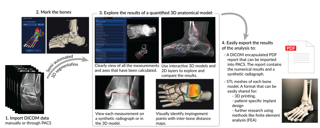
Comments are closed.

    Archives

    January 2022
    April 2021
    March 2021
    February 2021
    January 2021
    June 2020
    May 2020
    April 2020
    January 2020
    December 2019
    September 2019
    June 2019
    March 2019
    July 2018
    June 2018
    May 2018
    February 2018
    January 2018
    December 2017
    November 2017
    October 2017
    September 2017

    Categories

    All

    RSS Feed

Picture

Transforming Treatments

Are you interested in our products? Contact our sales team: sales@disior.com 
​

​
Do you need help?
Contact our support team: 
support@disior.com


Follow our journey at:

​
Copyright © 2023 Disior™ Ltd. All Rights Reserved Privacy Notice

License Agreement

Disior
™ a Paragon 28® Company

Picture
  • Home
  • Available Modules
    • Foot & Ankle
    • Planning
    • Hand & Wrist
    • CMF Orbital
  • Clinical Research
  • Expert Cases
  • News
    • Press
    • Blog
  • About us
    • Contact Us
    • Paragon 28®
    • Careers